ADOA

Autosomal Dominant Optic Atrophy
An image of a male toddler looking to his right and smiling. The toddler is surrounded by bubbles floating in the air.
DISEASE OVERVIEW

ADOA is a debilitating, blinding eye disease caused by insufficient gene expression of the OPA1 gene in optic nerve cells of the eye.

Patients experience progressive and irreversible vision loss, with many experiencing their first symptoms of vision loss before 10 years of age. There are no current treatments available for ADOA patients.

PYC has developed PYC-001, a potentially disease-modifying drug that is unique in its potential for full restoration of cell function. PYC-001 is currently in clinical studies in ADOA patients and is the first precision therapy to be dosed in patients with ADOA.

ADOA at a glance

An overview of key disease information.
An icon of a navy blue globe with an orange stand.

An urgent need to create treatments

ADOA affects 1 in every 35,000 people.1,2

An icon of two strands of RNA next to each other. The strand on the left is navy blue and the strand on the right is orange. Both strand are otherwise identical. They are both gently curved vertical lines with short horizontal lines staggered downwards like the rungs of a ladder.

ADOA is a monogenic disease

ADOA is caused by a mutation in one of the patient’s two copies of the OPA1 gene in the retinal ganglion cells (RGCs) that form the optic nerve of the eye.

An icon of a navy blue eye with an orange circle containing an exclamation mark in front of it, in the bottom right corner.

The median age of ADOA onset is 7 years of age

80% of patients experience symptoms before 10 years of age.1

An icon of a navy blue eye with an orange diagonal slash across the front of it from the top right corner to the bottom left corner.

Loss of central vision

Patients with ADOA initially experience central vision loss and then peripheral vision loss.

An icon of an orange circle containing the abstract PYC logo in white. There is a navy blue dotted arrow around the orange circle.

PYC’s drug candidate, PYC-001, seeks to stop disease progression

ADOA patients have a mutation in one copy of the OPA1 gene – this leads to their retinal cells producing half as much OPA1 protein as is required for normal function. PYC-001 seeks to restore the production of OPA1 protein in patients, therefore rescuing function of retinal cells and thus stopping disease progression.

An icon of a navy blue and orange clipboard with ticks on it next to a strand of orange DNA.

Encouraging results

PYC’s therapy, PYC-001, seeks to increase OPA1 protein expression in the RGCs to restore cell function and is currently in a Phase 1 clinical trial.

ADOA at the genetic level

Understand how a single genetic mutation causes lifelong disease.
An icon of four DNA spirals organised into two sets of pairs. The DNA pairs on the left are both blue and white, and are titled 'Unaffected DNA'. One DNA pair on the right is orange and white while the other is blue and white. This set of DNA is titled 'ADOA patient DNA'.

Genetic mutation

ADOA patients have a mutation in one of their two copies of the OPA1 gene – this mutation is present in all the cells in their body but it causes disease in the retinal ganglion cells (RGCs) in the optic nerve of the eye.

An icon of two sets of RNA pairs side-by-side. The set on the left is blue and is titled 'Unaffected person'. There is a white arrow below this pair pointing down towards two strands of OPA1 protein chains. On the right, one strand is orange and one is blue within the pair. It is titled 'ADOA patient'. There is a white arrow below this pair pointing down towards one strand of OPA1 protein chain and a white bubble containing the text '-50%'.

Decreased protein production

The OPA1 gene mutation creates an unstable RNA message – this leads to approximately half as much OPA1 protein production as is required to function normally. PYC’s therapeutic, PYC-001, seeks to increase protein expression in the remaining healthy copy of the OPA1 gene to restore OPA1 protein production to levels that are sufficient for the cells to function.

An sagittal cross-section infographic of an eye. The optic nerve is depicted in orange at the back of the eye. This is titled 'Optic nerve'.

Cellular changes

Insufficient production of the OPA1 protein causes RGCs to die – this leads to degeneration of the optic nerve. The optic nerve is responsible for sending visual messages from the eye to the brain.


PYC-001 has disease-modifying potential to halt cellular changes and arrest disease progression – it’s a potentially life-changing therapeutic.

An icon of a white eye with an orange diagonal slash through it.

Patient symptoms

As the number of RGCs in the optic nerve decline, ADOA patients experience changes in their vision, including central vision loss and eventually blindness.


At PYC, we aim to eradicate the development of ADOA symptoms with our first-in-class drug candidate, PYC-001.

The human impact of ADOA

ADOA leads to progressive and irreversible sight changes from childhood.
A photo of a tram on a busy street in a city.

10 years old

Patients are usually diagnosed in early childhood.

A photo of a tram on a busy street in a city. The image has decreased opacity and is difficult to make out, especially in the centre of the image.

30 years old

At first, patients lose their central vision and then their peripheral vision.

A photo of a tram on a busy street in a city. The image has a very low opacity and is difficult to see and the centre of the image is completely white.

50 years old

Patients are often declared legally blind at around 50 years of age, which significantly impacts their quality of life.

PYC-001 is a potentially life-changing treatment

PYC-001 is a revolutionary RNA therapeutic that increases OPA1 protein levels to enhance the mitochondrial structure and cellular function of disease-affected retinal ganglion cells (RGCs) in ADOA patients. PYC-001 is a breakthrough therapy designed to not only treat ADOA, but fully restore cell function in a mutation-independent manner.

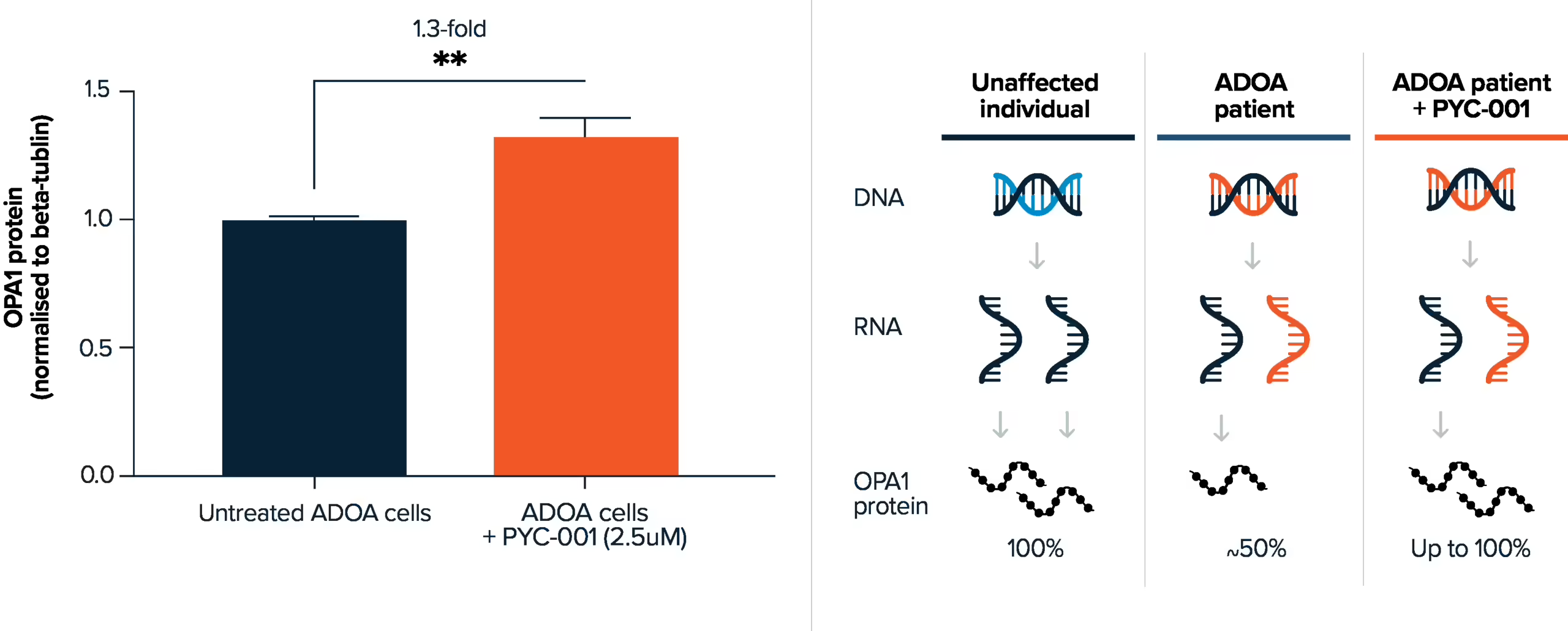
With recent advances in technology, PYC can grow RGCs of ADOA patients using patient-derived models and treat these patient-like cells with PYC-001 to demonstrate the efficacy of the RNA drug. Through increasing OPA1 gene expression, PYC-001 increases OPA1 protein production of ADOA RGCs in patient-derived models.

A graph and a table side-by-side. The y-axis of the bar graph on the left is titled 'OPA1 protein (normalised to beta-tubulin)'. There are two bars in the graph, the left bar is titled 'Untreated ADOA cells' and reaches a value on the y-axis of approximately 1.0. The right bar is titled 'ADOA cells + PYC-001 (2.5uM)' and reaches a value on the y-axis of approximately 1.3. Above the two bars there is a subtitle that reads '1.3-fold increase'. The table on the right contains three columns titled 'Unaffected individual', 'ADOA patient' and 'ADOA patient + PYC-001' from left to right. In the 'Unaffected individual' column there is light and dark blue DNA, two strands of dark blue RNA and two strands of OPA1 protein, these are labelled '100%'. In the 'ADOA patient' column there is dark blue and orange DNA, one strand of dark blue and one strand of orange RNA and one strand of OPA1 protein, labelled '~50%'. In the 'ADOA patient + PYC-001' column there is dark blue and orange DNA, one strand of dark blue and one strand of orange RNA and two strands of OPA1 protein, these are labelled 'up to 100%'.
Normalised fold-change in expression of OPA1 protein assessed by western blotting. Bar graph represents mean ± SEM @ day 7 PPMO incubation, Patient; OPA1 c. 287delAn=3 biological replicate, 3 technical replicates. Student's t-test. *=p<0.001.

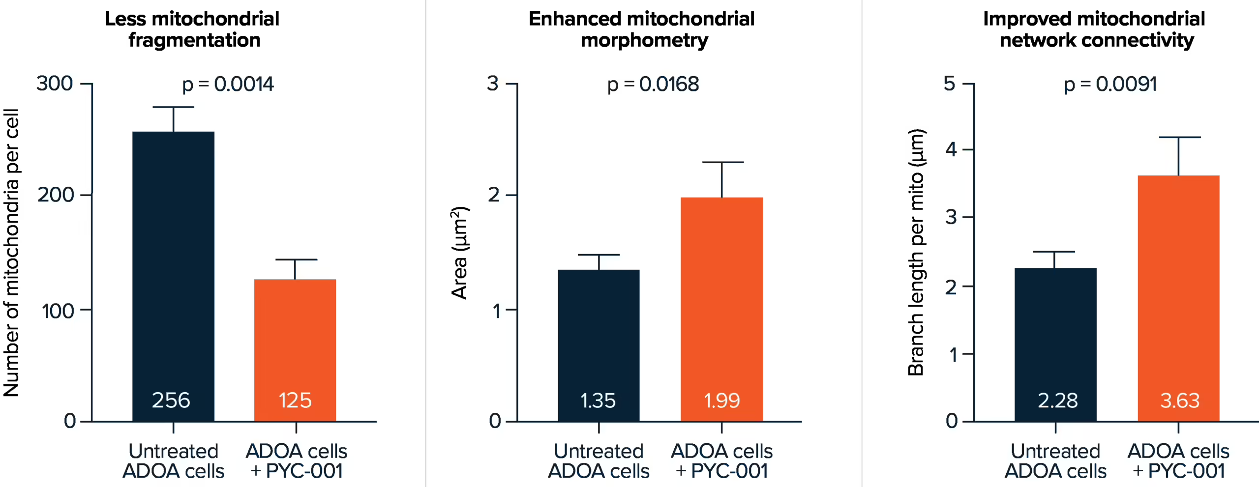
Pre-clinical studies have shown that PYC-001 increases OPA1 protein production back towards normal levels, resulting in enhanced RGC mitochondrial function that fully restores ADOA disease-affected cells.

Three bar graphs side-by-side. The graph on the left is titled 'Less mitochondrial fragmentation' and the y-axis is titled 'Number of mitochondria per cell'. There are two bars in this graph, the bar to the left is titled 'Untreated ADOA cells' and achieves a value of 256. The bar to the right is titled 'ADOA cells + PYC-001' and achieves a value of 125. There is a small label above the left bar that reads 'p=0.0014. The middle graph is titled 'Enhanced mitochondrial morphometry' and the y-axis is titled 'Area (um2)'. There are two bars in this graph, the bar to the left is titled 'Untreated ADOA cells' and achieves a value of 1.35. The bar to the right is titled 'ADOA cells + PYC-001' and achieves a value of 1.99. There is a small label above these bars that reads 'p = 0.0168'. The right graph is titled 'Improved mitochondrial network connectivity' and the y-axis is titled 'Branch length per mito (um)'. There are two bars in this graph, the bar to the right is titled 'Untreated ADOA cells' and achieves a value of 2.28. The bar to the left is titled 'ADOA cells + PYC-001' and achieves a value of 3.63. There is a small label above these bars that reads 'p=0.0091'.
Bar graph represents mean+SEM @ day 7 PPMO incubation, Patient 2 derived fibroblast harbouring OPA1 c.2708delTTAG mutation, n=1 biological replicate, 1 technical replicate. Student's test.

Patients are our priority

An image of a female healthcare worker wearing a white coat and stethoscope who is seated at a desk. She has a pen and clipboard in hand and is smiling as she talks to a woman in casual clothes who is seated across from her.

Our commitment to patients

We believe it is essential to understand the people living with the genetic diseases that we are aiming to treat. At PYC, we make it a priority to collaborate with patients and caregivers across our development process – to ensure that our RNA therapeutics best meet their needs.

An image of a person in a white coat sitting down and holding a pen in their right hand and a clipboard in their left. In the background there is a person in plain clothes sitting next to them.

Our patient trials

At PYC, we conduct responsible patient trials to ensure that our treatments are safe and effective. These trials rely on patient volunteers and are crucial to the development of disease-modifying therapies for genetic diseases.

References

  1. Yu-Wai-Man P, Griffiths PG, Burke A et al. The prevalence and natural history of dominant optic atrophy due to OPA1 mutations. Ophthalmology. 2010 Aug;117(8):1538-46, 1546.e1. doi: 10.1016/j.ophtha.2009.12.038. ↩︎
  2. Amati-Bonneau P, Milea D, Bonneau D et al. OPA1-associated disorders: phenotypes and pathophysiology. Int J Biochem Cell Biol. 2009 Oct;41(10):1855-65. doi: 10.1016/j.biocel.2009.04.012. ↩︎
SUBSCRIBE

Keep up with all the latest changes

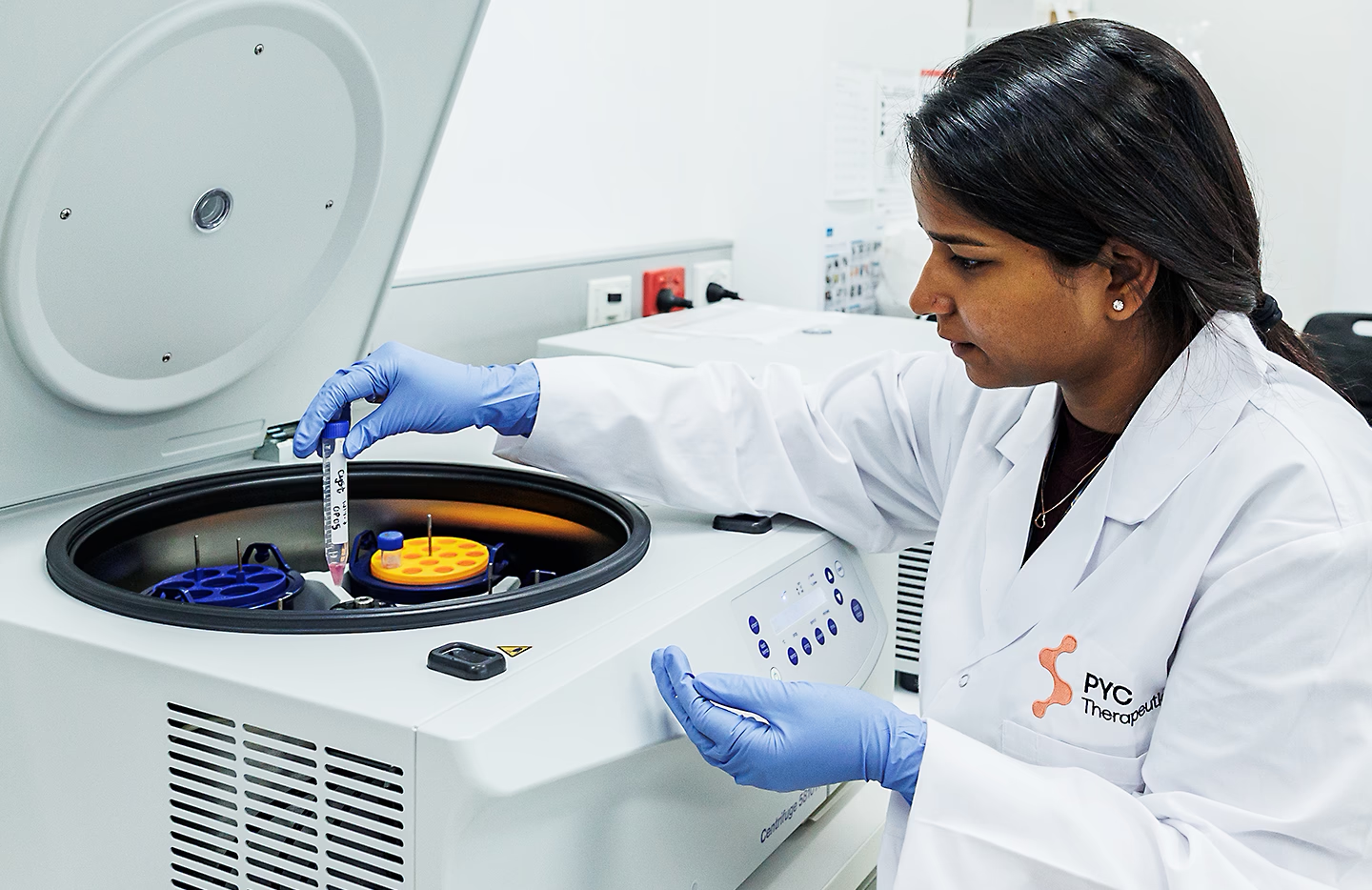
    A photo of a female scientist wearing a white lab coat and blue gloves in a lab. She is placing a test tube inside a centrifuge.